

You know exactly which move sets it off. You’ve stopped mentioning it because the tests come back fine and the treatments don’t stick.
Part of you has started to wonder if this is just what your body does now.
It isn’t aging. It may be one small joint that no one has checked.
Most adults over 50 with this problem are managing the wrong diagnosis. They treat general back strain. An unstable or inflamed SI joint keeps deteriorating without targeted care.
The sacroiliac joint connects your spine to your pelvis. It is one of the most common sources of chronic low back pain in people your age.
It is also one of the most commonly missed.
This article is for informational purposes only and does not constitute medical advice. SI joint pain shares symptoms with other conditions and requires a proper clinical evaluation. Always consult a qualified healthcare provider before making any changes to your treatment or care.
Three Moves That Load the SI Joint and What Happens When One Does
You already know this pattern. SI joint back pain doesn’t spread evenly. It catches you during specific moves, in a specific spot, every time.
That isn’t a mystery. That’s a clue.
The SI joint accounts for 15 to 30 percent of cases of chronic, nonradicular low back pain.¹
Between one in six and one in three people with persistent back pain have this joint as the actual source.
Not a disc. Not a lumbar nerve.
The three moves below are structurally predictable. Each one places shear, torsion, or uneven compression on the sacroiliac joint. Healthy tissue handles these forces. Inflamed or unstable tissue does not.
THE 3 MOVES
- Rising from a seated position
Shifts full weight through one hip at a time, placing uneven load across the joint. - Rolling over in bed
Rotates the pelvis against a fixed sacrum, stressing the posterior ligaments. - Single-leg loading
Stair climbing or putting on a shoe. The joint must stabilize one side of the pelvis while the other leg moves.
If any of these reproduce your pain in the same spot every time, write it down.

The pain is typically one-sided. It sits just below your beltline, often in the upper buttock.
It may radiate down the back of your thigh. That happens because of the SI joint’s referral pattern, not because of a compressed nerve.
Most people with this pain have been told it’s a muscle strain or a disc problem. Both diagnoses keep the SI joint off the table entirely.
That’s a problem worth understanding. A muscle strain resolves with rest. A disc issue responds to traction or decompression.
Neither approach targets a joint that can’t share load correctly. If the diagnosis is wrong, the treatment misses the source.
The pain keeps coming back because the actual structure is never addressed.
If those three moves are that predictable, the structure causing them has a name.
Why Your Body Stopped Warning You Gradually and Started Punishing You Specifically
There was a before and an after. A point where the pain stopped being background stiffness. It became a specific event you can predict to the second.
That shift reflects something real in a specific structure.
A CT imaging study found some degenerative change in the sacroiliac joints of every symptom-free adult aged 50 or older it examined.² After 50, wear exists in this joint for everyone.
The question isn’t whether wear is there. The question is whether the joint can still absorb load without triggering pain.
When it can’t, pain becomes specific. It follows the same move, in the same joint, because that joint is the one no longer managing the force.
Think of it this way. A chair leg that is cracked holds fine until you sit down hard. The crack was always there. The load revealed it.

Your SI joint works the same way. The degenerative change builds silently for years. A specific movement finally makes the stressed tissue announce itself.
That specificity isn’t a sign something is beyond repair. It’s a sign a particular structure needs attention.
Most people at this point increase stretching or rest more. Both can help with muscular soreness. Neither targets a joint that has lost the ability to share load correctly.
Most low back workups never give this joint attention. The reason is worth knowing.
The SI Joint Has One Job, and After 50 That Job Gets Harder
The sacroiliac joint sits on each side of your lower back. It’s where your spine meets your pelvis. It’s also the largest axial joint in your body.
Axial means it runs along the central axis of your skeleton.
Its job is load transfer. Every step sends your upper body’s weight through this joint and down to your legs.
It absorbs shear, compression, and torsion at the same time, with every stride.
The joint barely moves. Its rotations do not exceed about three degrees in any plane. Yet it transfers large bending forces and compression loads continuously.³
Small motion. Enormous job.
Two systems keep it stable. Ligaments act as passive restraints. They keep the joint within a safe movement range.
The gluteus maximus, pelvic floor, and deep abdominals act as active stabilizers. They fire around the joint to manage forces the ligaments can’t handle alone.
After 50, both systems are under pressure. Ligament tissue loses tensile strength with age. The stabilizing muscles weaken if they aren’t specifically trained.
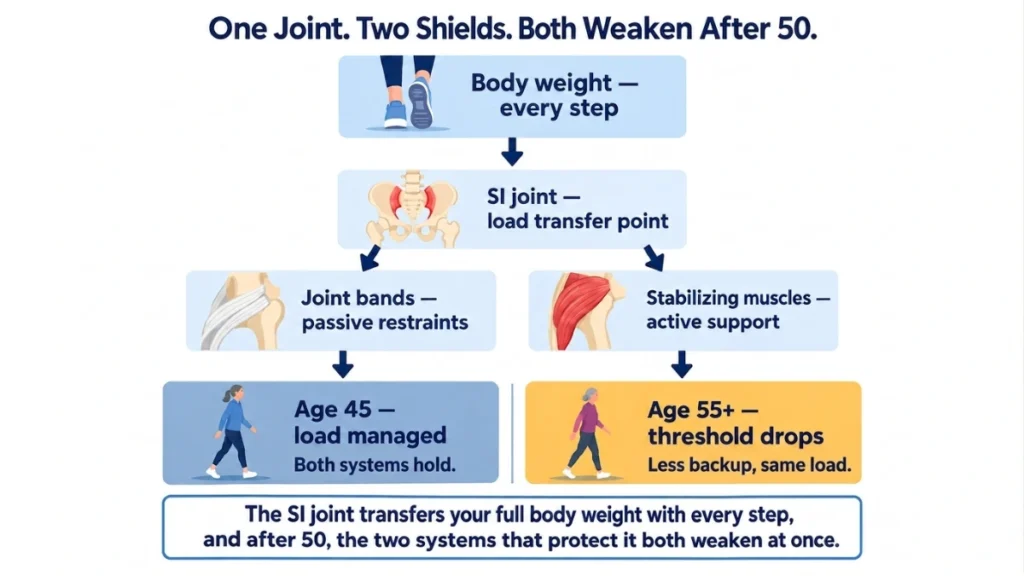
When both drop, the joint takes the same loads with less backup. The math doesn’t work in the joint’s favor.
Research doesn’t point to one single failure. What it shows is that the threshold for pain gets lower as support decreases.
A load the joint managed at 45 may be enough to provoke it at 55.
What gives way first?
Why Most Low Back Diagnoses Miss the SI Joint Entirely
Most imaging ordered for low back pain doesn’t include the SI joint. Standard MRI and X-ray scans focus on the lumbar vertebrae and the discs between them.
The SI joint isn’t in frame.
Here’s the contradiction. This joint accounts for 15 to 30 percent of chronic, nonradicular low back pain.¹ The tools to identify it exist. The physical tests take minutes to perform.
A 2013 review by Cohen et al. in Expert Review of Neurotherapeutics stated: “Although no single physical examination or historical feature can reliably identify a painful SIJ.”¹
That’s the honest clinical picture. No single test confirms it alone. A combination does.
Part of the problem is that pain from this joint mimics other conditions. The referral pattern overlaps with sciatica. The location overlaps with lumbar disc pain.
The history sounds like hip arthritis. Without a targeted exam, the joint stays invisible.
Five standard tests assess sacroiliac joint dysfunction directly:
- FABER (hip flexed, abducted, externally rotated)
- Thigh thrust
- Compression
- Distraction
- Gaenslen’s test
Three or more positive results from the five-test battery of FABER, compression, distraction, thigh thrust and Gaenslen produce 91 percent sensitivity and 78 percent specificity.⁴
In people who do have SI joint pain, three positive tests out of five catch it about 91 percent of the time.
Those tests take minutes in a clinic. The wait to reach that clinic can take years.
What Happens Inside the Joint When Those Three Moves Repeat Without Correction
Pain receptors live inside the SI joint. Not just around it.
The joint capsule, the ligaments, and the bone just beneath the cartilage all contain nociceptors.⁵
Nociceptors are the nerve endings that send pain signals to your brain.
They are present throughout the joint. The capsule, ligaments, and subchondral bone all carry them.
When these tissues are inflamed and then repeatedly loaded, the signal comes from inside the joint itself. This isn’t a nerve problem nearby. It’s the joint tissue firing directly.
Each time an inflamed joint absorbs an unmanaged load, it adds a small insult to already irritated tissue. The capsule, ligaments, and bone beneath the cartilage absorb it again.
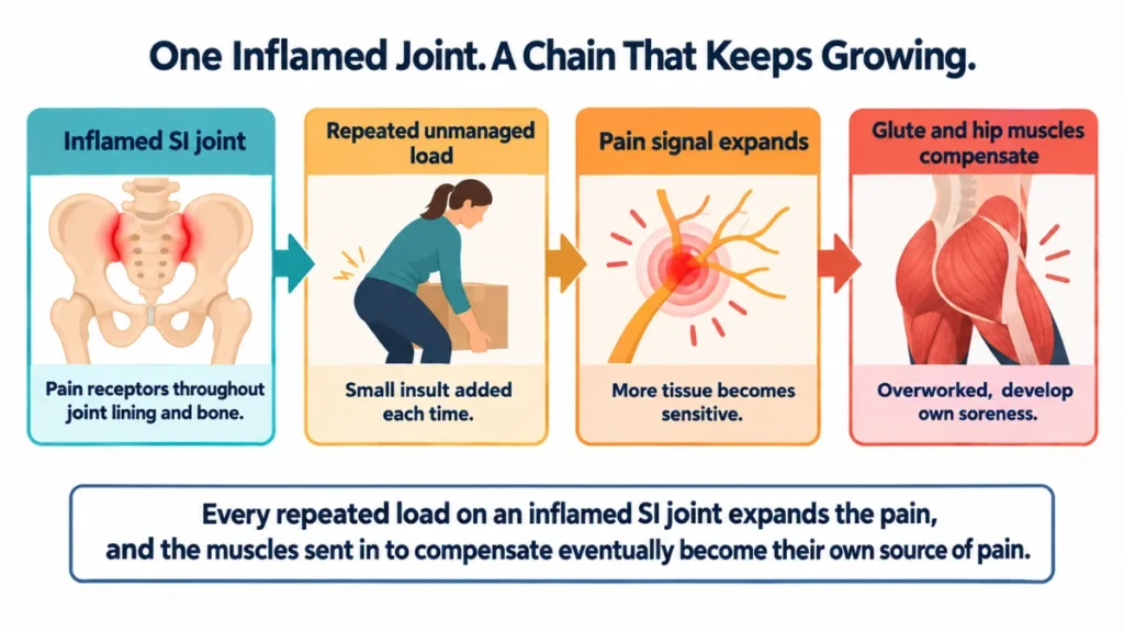
Over time, the pain expands. What started as one predictable trigger can spread as more tissue becomes sensitized.
Surrounding muscles tighten to protect the joint. That compensation creates its own pain pattern.
The glutes and hip flexors begin managing load the joint should be sharing. They weren’t built to do this alone. Over months, the overworked muscles develop their own soreness.
By that point, many people are treating the muscle pain and ignoring the joint that started it. The original source becomes harder to isolate.
The pattern can be redirected. But the longer it runs, the harder that becomes.
How to Bring What You Just Learned Into a Clinical Conversation
You now have something most patients don’t: a specific movement, a specific location, and a name for what may be happening.
Tell a clinician this. “The pain is on one side, below my beltline.”
“It happens consistently when I [name the move]. I’ve tracked which movements reproduce it. I’d like the sacroiliac joint assessed.”
That language is specific enough to move the appointment forward.
Physical therapy is the standard first step. It should target sacroiliac joint mechanics directly.
A program focused on the gluteus maximus and pelvic stabilizers addresses the active support system the joint relies on.

Exercise and manipulation therapy can improve pain and disability from this condition. Benefits have been documented for up to 24 weeks.⁵
If physical therapy isn’t enough, the next step is a fluoroscopy-guided diagnostic injection. Local anesthetic goes directly into the joint.
If the pain disappears, the joint is confirmed as the source.
After that, radiofrequency ablation can interrupt the nerve signal from the joint. Surgical fusion is reserved for cases where all other options have failed.
The treatment path exists. Getting on it requires naming the right joint first.
Every month spent managing a lumbar problem that isn’t there is a month this joint goes without care.
The longer the surrounding muscles compensate for an untreated joint, the more the pattern embeds itself.
Catching this early matters. That’s the risk in waiting.
What Acting on This Gets You
Targeting the right joint means the right treatment. Test the three flagged movements at home, identify whether each one reproduces the pain pattern described, and bring specific findings to a physical therapist or physician for SI joint assessment. SI joint back pain is manageable. The first step is knowing what you’re dealing with.
References
- Cohen SP, Chen Y, Neufeld NJ. Sacroiliac joint pain: a comprehensive review of epidemiology, diagnosis and treatment. Expert Review of Neurotherapeutics. 2013;13(1):99-116. https://www.tandfonline.com/doi/full/10.1586/ern.12.148
- Shibata Y, Shirai Y, Miyamoto M. The aging process in the sacroiliac joint: helical computed tomography analysis. Journal of Orthopaedic Science. 2002;7(1):12-18. https://www.sciencedirect.com/science/article/abs/pii/S0949265815332437
- Kiapour A, Joukar A, Elgafy H, Erbulut DU, Agarwal AK, Goel VK. Biomechanics of the Sacroiliac Joint: Anatomy, Function, Biomechanics, Sexual Dimorphism, and Causes of Pain. International Journal of Spine Surgery. 2020;14(s1):S3-S13. https://www.ijssurgery.com/content/14/s1/S3
- Buchanan P, Vodapally S, Lee DW, Hagedorn JM, Bovinet C, Strand N, Sayed D, Deer T. Successful Diagnosis of Sacroiliac Joint Dysfunction. Journal of Pain Research. 2021;14:3135-3143. https://doi.org/10.2147/JPR.S327351
- Aranke M, McCrudy G, Rooney K, Patel K, Lee CA, Hasoon J, Urits I, Viswanath O, Kaye AD. Minimally Invasive and Conservative Interventions for the Treatment of Sacroiliac Joint Pain: A Review of Recent Literature. Orthopedic Reviews. 2022;14(2). https://orthopedicreviews.openmedicalpublishing.org/article/31915-minimally-invasive-and-conservative-interventions-for-the-treatment-of-sacroiliac-joint-pain-a-review-of-recent-literature