

You sat in the office while the doctor scrolled through your results. TSH: normal. T4: normal. “Everything looks fine,” they said. And yet you are reading slower than you did two years ago, losing words mid-sentence, and watching the sharpness you relied on quietly recede. You have already been told it is probably stress, or age, or not sleeping well enough. You have tried all three explanations on for size.
For millions of adults over 50 who feel mentally slower than they used to, the standard thyroid panel was not designed to measure what thyroid hormone is actually doing inside the brain. Thyroid and brain fog are more connected than most lab reports suggest, and that connection runs through biology your blood test does not reach.
Why Read This
- Why your brain maintains its own thyroid hormone supply that no blood test measures, and what happens when that supply runs short
- The specific thinking tasks that go quiet first, often years before any lab number moves
- A gene variant that makes standard treatment work less effectively in the brain while TSH reads completely normal
- What a more complete thyroid picture includes, and how to bring a documented case to your next visit
This article is for informational purposes only and does not constitute medical advice. If you are experiencing cognitive symptoms or have concerns about your thyroid health, speak with a qualified healthcare provider before making any changes to your testing or treatment.
What thyroid hormone actually does inside a brain cell
If you have ever wondered why clearing up a thyroid problem does not always clear up the foggy thinking that came with it, the answer starts here, at the level of a single brain cell.
Your brain does not just receive thyroid hormone from the bloodstream and use it directly. It runs its own conversion system. The inactive form, thyroxine [the hormone the thyroid gland mostly releases, also called T4], travels through the blood and crosses into the brain.
Once inside, T4 reaches specialised support cells called astrocytes [star-shaped brain cells that feed and protect neurons].
Astrocytes carry an enzyme called type 2 deiodinase [an enzyme that converts the inactive thyroid prohormone T4 into the active form T3] that strips one atom from T4 and converts it into the active form, triiodothyronine [the active thyroid hormone that neurons actually use, also called T3]. That T3 is then passed to the neurons next door, which use it to regulate gene expression, maintain signal speed, and support the processes behind memory and attention.
Here is what this means for understanding thyroid and brain fog: your brain’s T3 supply comes from two sources. Roughly half arrives from general circulation, and the other half is manufactured locally inside astrocytes from the T4 your blood carries there.¹
The two supplies are partially independent. Circulating T3 levels can look fine while the local conversion system runs short, and no standard blood panel sees the local system at all.
Many people who still feel cognitively off after a thyroid diagnosis have been told their numbers are fine and their symptoms must come from somewhere else. That experience has a physiological basis.
It begins in the gap between what the blood test measures and what the brain’s own hormone system is doing.
This system does most of its known work in animal models and in early human tissue studies. Whether the exact conversion proportions hold identically in adult human brains remains an open question.¹
The basic architecture, the two-source T3 supply and the astrocyte conversion step, is well established across the research.
What the blood test tells your doctor and what is happening at the neuron are two different readings, and the gap between them is where the cognitive story begins.
Why your TSH number can look fine while your thinking slows down
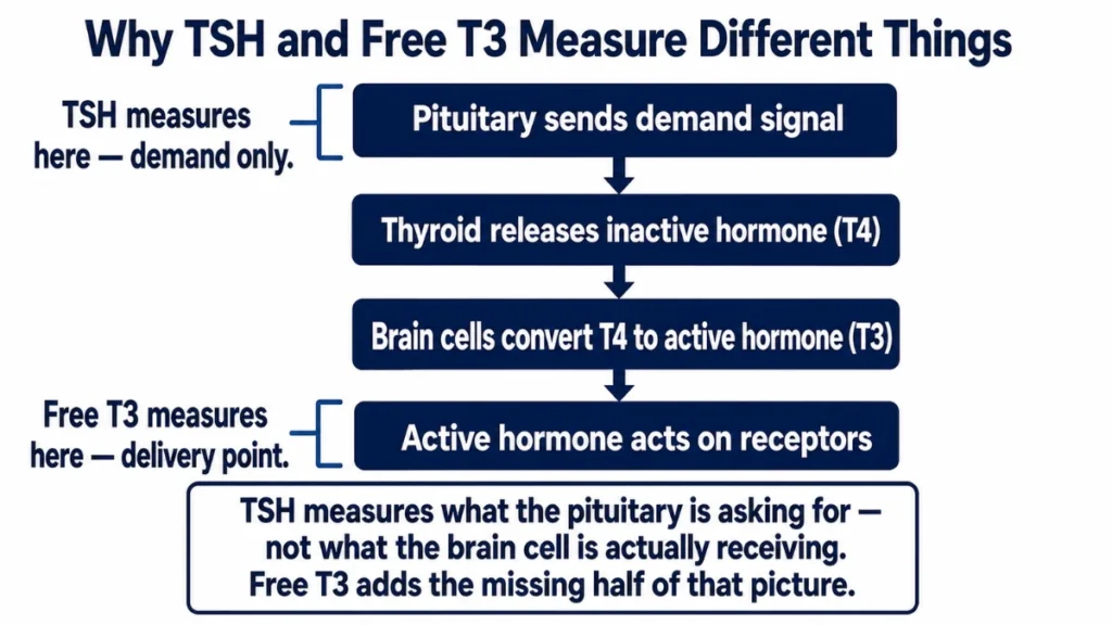
When your doctor says your TSH is normal, what that result is actually measuring may not be what you think. TSH is not a direct reading of hormone levels. It measures how loudly the pituitary gland is signalling the thyroid to make more. When the pituitary senses that circulating hormone levels are adequate, it quiets that signal.
A normal TSH means the pituitary is satisfied. It says nothing about what thyroid hormone is doing once it arrives at a brain cell.
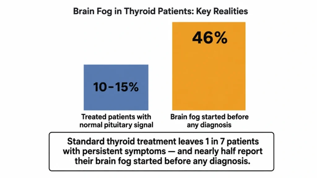
The TSH test measures what your pituitary is asking your thyroid to do, not what thyroid hormone is doing inside your brain cells, and those are not the same measurement.
That gap shows up in the data. Among people treated for hypothyroidism whose TSH has been brought to normal levels, research suggests that roughly 10 to 15 percent continue to experience persistent symptoms, including fatigue, low mood, and cognitive difficulty.²
Those symptoms are not imaginary and they are not explained by the TSH number, because the TSH number never addressed the brain’s local hormone system in the first place.
Many people’s experience of cognitive slowing actually predates the point at which their thyroid problem became detectable. In a large patient survey, 46 percent of people who reported brain fog while being treated for hypothyroidism said their symptoms had begun before their hypothyroidism was diagnosed.² Most had normal TSH at that time.
Blood panels can read completely normal while the brain runs low on active thyroid hormone.
In animal studies, mice carrying a common DIO2 gene variant had normal circulating TSH, T4, and T3, while their brains were measurably hypothyroid.³
Human replication is ongoing, but the pattern illustrates what the standard panel was not designed to detect.
No single study directly proves in adult humans that thyroid dysfunction causes cognitive symptoms while TSH stays normal. The case is assembled from multiple lines of evidence, each testing a different piece.
The brain’s local T3 system is established in animal and tissue research. The persistent symptom data comes from treated patient surveys. The gene variant work is from cell lines and mice.
Together they build a coherent explanation, but the full chain has not been tested in one controlled human trial.

If your doctor has told you a normal TSH means your thyroid is not the problem, that statement is accurate at the level of the pituitary signal. It is incomplete at the level of the brain cell.
Knowing which cognitive tasks go quiet first, and in what order, is what the next section covers.
The cognitive tasks that go quiet before any test catches them
You notice it in specific moments rather than as a general fog. The word you need is there, and then it is not. A name, a number, the next item on a task you have done a hundred times. These are not random lapses. They follow a pattern that research on hypothyroidism has documented across multiple neuropsychological studies.
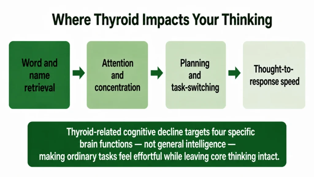
Word retrieval is usually the first specific symptom people can name: the word that won’t come, the sentence that stalls mid-thought.
Research on hypothyroid states consistently finds that memory [the brain’s ability to store and retrieve words, names, and sequences on demand] is the most affected domain, with verbal memory producing the clearest and most reproducible deficits.⁴
Beyond memory, attention and concentration also show measurable decline in hypothyroid states, along with executive function [the mental processes that allow planning, switching between tasks, and holding multiple pieces of information at once].⁴ Psychomotor function [the speed at which a thought translates into a physical or verbal response] rounds out the affected profile.
Taken together, these findings point to a pattern: the systems most dependent on reliable hormone signalling for speed and retrieval are the ones that slip first.
Global intelligence, the broad measure most people think of as “being smart,” tends to be spared or minimally affected. The deficit is narrower than that. It is the retrieval speed, the switching cost, the word that won’t come, that shows up first.

Thyroid-related cognitive change is not a drift toward stupidity. It is a narrowing of specific systems, verbal retrieval above all, that leaves general intelligence intact while making ordinary tasks feel effortful. That is a precise biological signature, not a vague complaint.
Distinguishing thyroid-related cognitive change from other causes is genuinely difficult, because the profile overlaps with normal ageing, sleep disruption, and depression.⁴ The symptoms do not arrive labelled.
Determining whether the source is hormone-related rather than something else is one of the harder questions in this space, and the profile alone cannot answer it.
The gene variant that makes standard thyroid treatment work differently for some people
Some people do everything their doctor recommends for a thyroid condition. Their TSH normalises. Their levels look right. And they still feel exactly the cognitive flatness the previous section describes. One biological reason for that persistence sits in a gene variant that a large portion of the population carries without knowing it.
DIO2 [the gene that encodes the type 2 deiodinase enzyme responsible for converting T4 into active T3 inside cells] is the relevant gene. A common variant of this gene is called Thr92Ala. It changes the deiodinase enzyme’s structure in a way that is associated with endoplasmic reticulum stress [a state in which a cell’s protein-folding machinery is overwhelmed, reducing the enzyme’s output] in glial cells.³
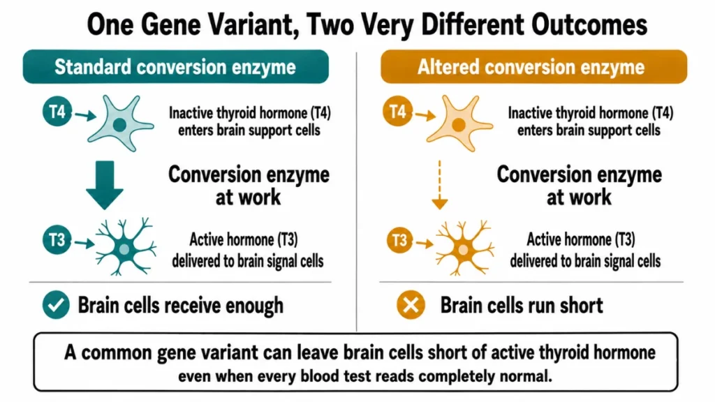
Cell studies and mouse models show that the Ala92 version of the enzyme may produce approximately 30 percent less active T3 from T4 over 24 hours than the standard version.³
This research was conducted in HEK-293 cells and in genetically modified mice, not in human subjects. The study found that Ala92-Dio2 mice showed brain hypothyroidism while their circulating TSH, T4, and T3 all remained normal.³
Whether the same mechanism operates at the same scale in human brains is not yet confirmed, and the research authors explicitly called for human clinical studies to follow.
Standard treatment looks different through the lens of this variant. Levothyroxine, the most common thyroid medication, provides T4. The body and brain then convert that T4 to T3.
If the DIO2 enzyme is less efficient at that conversion, supplementing T4 alone may not adequately supply T3 to brain cells, even when blood tests suggest all levels are normal. The Ala92-Dio2 mice in the study did not respond to levothyroxine treatment the way mice without the variant did.³

Carrying the Thr92Ala variant does not automatically produce symptoms. The association between the variant and cognitive symptoms is observed in some studies but not confirmed in others.³ Research in this area is still developing.
Clinical evidence connecting this gene variant to outcomes in human hypothyroid patients remains limited and inconsistent. Not every person with the variant has a problem, and current evidence does not support routine testing for it as part of standard thyroid care.
What it does support is the principle that standard treatment normalises a blood number, not necessarily brain cell function.
A routine panel has limits. A more complete one might at least approach what it cannot answer.
What a fuller thyroid picture actually includes
Feeling cognitively slow after treatment for a thyroid condition may mean you are working with an incomplete picture of your own hormone status. A growing number of clinicians argue that TSH alone tells only part of the story, and that free T3 and free T4 measured together provide a better view of what thyroid hormone is actually doing in the body.
At its core, TSH is a pituitary hormone, not a thyroid hormone. It reflects the demand side of the signal chain, not the delivery side.
A patient whose TSH sits at 2.0 and whose free T3 sits near the low end of the reference range has less active hormone available at the receptor than a patient with the same TSH and a mid-range free T3. The blood T3 level, even an imperfect proxy for brain T3, adds a piece of information that TSH cannot.⁵
Thyroid peroxidase antibodies [proteins the immune system produces that target the gland itself, commonly elevated in Hashimoto’s thyroiditis] are another marker some clinicians include.⁷ When antibody levels are elevated, immune activity against the thyroid is already underway, often before the gland has lost enough function to move the TSH number.
Evidence for free T3 as a clinical guide is thinner than the clinical interest in it. The case rests primarily on practitioner observations, retrospective patient data, and arguments from clinicians who find the TSH-alone approach insufficient.
There is no large, long-term, randomised trial that has proven that treating to a free T3 target improves cognitive outcomes better than standard TSH-based care. The source most directly making this case is a single-author perspective piece that carries a published correction.⁵
This is where the field is, not where it has landed.

Practical guidance is still possible in that context. If someone is being treated for a thyroid condition and still experiences cognitive symptoms despite a normal TSH, the conversation with their doctor about testing free T3 and antibody levels is reasonable and supported by the weight of the clinical discussion, even where the controlled trial evidence does not yet exist.
The recommendation may apply differently depending on the individual’s treatment history, medication type, and other health factors.
A more complete panel raises better questions. Getting the most from it depends on what you track before you walk in.
What to notice, track, and bring to your next appointment
Most people walk into a thyroid appointment with a feeling and walk out with a number. The number either explains the feeling or it does not, and if it does not, the visit is effectively over. Coming in with a documented pattern changes that dynamic.
Before the appointment, track your cognitive symptoms for three to four weeks: not a diary of complaints, but a specific log. Note when your thinking feels slowest, what tasks are hardest, how sleep and energy interact with it, and whether anything makes it better or worse on a given day.
Symptoms can persist even after TSH normalises. A clinical study found that treated hypothyroid patients, with a group mean TSH of 2.16, still scored measurably higher on symptom burden than euthyroid controls.⁶
A documented symptom pattern, rather than a general complaint, gives that conversation specificity.
When you go in, the questions worth raising with your doctor are: Has my free T3 been tested alongside TSH? If so, where does it sit within the reference range, not just whether it is technically inside it? Are thyroid peroxidase antibodies part of my picture?
Talk to your doctor before requesting or changing any testing or treatment plan, particularly if you are managing other conditions or taking medications that interact with thyroid function.

Cognitive symptom tracking log: what to record each day for three to four weeks
- Time of day when thinking feels slowest or sharpest
- Specific tasks that felt difficult: word retrieval, following a conversation, switching tasks
- Sleep quality the night before, rated simply (good, broken, poor)
- Energy level, mid-morning and mid-afternoon
- Any notable changes in stress, food, physical activity, or daily routine
- Anything that seemed to help or worsen mental clarity that day
Bring the log. Ask for the numbers, not just the verdict. A doctor who can see a pattern over weeks, matched against lab values, is in a better position to assess whether the picture is complete.
Accessible background on these conditions and testing options is available at thyroid.org, through the American Thyroid Association’s patient resources.
What the log cannot do is diagnose. It can make the conversation more specific, and a more specific conversation is more likely to go somewhere useful.
Your Thyroid, Your Brain, Your Next Question
The standard thyroid panel was built for one purpose: detecting glandular failure. It was not built to measure what thyroid hormone does inside the brain’s own conversion system. That gap is real, and it explains why some people feel cognitively slow despite numbers that look fine.
Ask your doctor to test free T3 alongside TSH at your next thyroid check.
Science here is still developing, and a normal TSH is not meaningless. The question worth asking is not whether something is wrong, but whether the picture is complete.
References
- Morte B, Bernal J. Thyroid Hormone Action: Astrocyte-Neuron Communication. Front Endocrinol (Lausanne). 2014. https://www.ncbi.nlm.nih.gov/pmc/articles/PMC4038973/
- Samuels MH, Bernstein LJ. Brain Fog in Hypothyroidism: What Is It, How Is It Measured, and What Can Be Done About It. Thyroid. 2022. https://pmc.ncbi.nlm.nih.gov/articles/PMC9469742
- Jo S, et al. Type 2 deiodinase polymorphism causes ER stress and hypothyroidism in the brain. J Clin Invest. 2018. https://pmc.ncbi.nlm.nih.gov/articles/PMC6307951
- Samuels MH. Psychiatric and cognitive manifestations of hypothyroidism. Curr Opin Endocrinol Diabetes Obes. 2014. https://pmc.ncbi.nlm.nih.gov/articles/PMC4264616/
- Lindner HH. Clinical thyroidology: beyond the 1970s’ TSH-T4 Paradigm. Front Endocrinol (Lausanne). 2025. https://www.ncbi.nlm.nih.gov/pmc/articles/PMC12234311/
- Brokhin M, Danzi S, Klein I. Assessment of the Adequacy of Thyroid Hormone Replacement Therapy in Hypothyroidism. Front Endocrinol (Lausanne). 2019. https://www.ncbi.nlm.nih.gov/pmc/articles/PMC6763555/
- Kaur J, Jialal I. Hashimoto Thyroiditis. StatPearls. Updated February 6, 2026. https://www.ncbi.nlm.nih.gov/books/NBK459262/