

You wake up at 3 a.m. with your heart still pounding from a dream that felt like a movie. For adults over 50 who have vivid or active dreams and want to know what they mean for brain health, that moment deserves more than a shrug.
REM sleep is now understood as a neurological signal: not just a dream factory, but an observable window into brainstem health.
Most people treat vivid REM-stage dreams as curiosities, not realizing the same sleep stage carries one of the strongest early signals of brain disease yet identified. Understanding what the brain actually does during those dreams may change how you think about the night entirely.
Why keep reading:
- Find out which brainstem circuit produces your dreams and why its early damage shows up in sleep first
- Learn why some nighttime movements are a medical observation, not a quirky habit
- Discover what a protein starting in the gut has to do with your REM sleep
- See what decades of follow-up research reveal about how far in advance sleep behavior can signal change
- Understand exactly what to tell your doctor, and when
What your brain actually does during those vivid REM dreams
You have probably noticed that your most intense dreams come just before you wake up: that rush of images, voices, and movement that feels more like living than sleeping.
That experience belongs to REM sleep [rapid eye movement sleep, the stage where brain activity surges close to waking levels while your body stays still].
In younger adults, REM sleep makes up a substantial portion of the night, though the percentage varies between individuals.¹
What the brain does during this phase is genuinely unusual. Your eyes flicker behind closed lids. Your heart rate and breathing shift rapidly. The brain’s emotional center, the amygdala [a small, almond-shaped structure that processes fear and emotional memory], fires at near-waking intensity.²
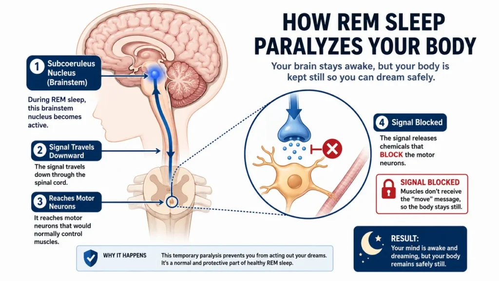
At the same time, your brainstem sends a chemical signal that paralyzes your skeletal muscles, a system called REM atonia [the temporary muscle paralysis that keeps you still while you dream].
REM sleep plays a central role in processing emotional experiences and consolidating emotional memories.²
Studies show this processing depends on active dreaming: people who recall their dreams overnight show measurable reductions in emotional reactivity the next morning, while those who do not report dreaming show no such effect.³
Emotional memory work is not the only function running. REM sleep preferentially preserves emotionally meaningful memories at the cost of neutral ones, a tradeoff that appears to serve long-term memory efficiency rather than pure retention.³
The brainstem machinery producing that paralysis matters enormously. It turns out to be the same hardware that fails first in a specific and common category of neurological disease.
Why losing muscle control in sleep is a warning worth understanding
If your bed partner has ever nudged you awake and said “you were punching in your sleep again,” you probably laughed it off. Most people do. It feels harmless: a vivid dream, a little movement, nothing broken. But that observation deserves a different kind of attention.

During healthy REM sleep, your brainstem enforces near-complete stillness. The moment a dream begins, signals travel from a brainstem region called the subcoeruleus nucleus downward through the spinal cord to block motor neurons from firing.⁴ Your muscles receive the command to stay quiet. This is REM atonia working as designed.
When atonia fails, the result has a name: REM sleep behavior disorder [a parasomnia in which the brainstem’s paralysis system breaks down, allowing people to physically act out their dreams].
People with this condition kick, punch, shout, and sometimes fall out of bed while still fully asleep.⁴ They are not sleepwalking. They are re-enacting their dreams in real time.
The same brainstem circuits that paralyze your muscles during REM sleep are among the first structures damaged when alpha-synuclein, the protein strongly linked to Parkinson’s disease, begins to misfold: your dreams are produced by the same hardware that breaks down earliest in that disease.⁵
That is not a theoretical concern. Between 70 and 91 percent of people diagnosed with isolated REM sleep behavior disorder will eventually develop Parkinson’s disease, dementia with Lewy bodies, or multiple system atrophy.⁵,⁴
The brainstem fails first. Dreaming shows the damage.
The protein that may start in your gut before it reaches your brain
You may have read about gut health and brain health as separate topics. They are not, and that connection is key to understanding where brainstem damage actually begins.
Alpha-synuclein [a protein that, when misfolded, forms toxic clumps called Lewy bodies that damage and kill neurons] is found throughout the nervous system, including the enteric nervous system [the web of nerve cells lining the gut wall, sometimes called the second brain].
A current hypothesis, supported by clinical and neuropathological evidence, holds that misfolded alpha-synuclein begins accumulating in the gut’s nerve network and travels upward through the vagus nerve toward the brainstem region governing REM atonia, before it ever reaches the dopamine-producing cells that motor symptoms damage.⁶
The Gut-to-Brain Pathway
The suspected 12-year progression of misfolded alpha-synuclein protein before neurodegeneration is fully diagnosed.
The Gut Network
Proteins begin misfolding and accumulating in the enteric nervous system.
The Vagus Nerve
Misfolded proteins use this major nerve as a highway, traveling upward.
The Brainstem
Damage reaches the subcoeruleus nucleus, breaking the sleep paralysis system.
The Motor Center
The progression finally damages the dopamine-producing cells.
Animal research injecting alpha-synuclein directly into the gut has reproduced this progression, showing REM-atonia disruption appearing before motor symptoms in the same temporal sequence.⁷ Human application of this animal finding has not been confirmed by a clinical trial.
The full picture is assembled from multiple research teams testing different pieces, rather than from a single study that directly proves the chain.
This matters because constipation and reduced sense of smell often appear in the same patients who later develop REM sleep behavior disorder, sometimes years before any movement problem is noticed.⁶ These symptoms make sense if the gut and olfactory nerves are early staging areas for the same protein.
Prodromal symptoms [early signs that appear before the defining features of a disease are clinically visible] like these are why researchers now treat isolated REM sleep behavior disorder as an event in a longer biological story, not a standalone sleep complaint.
Are you awake to what that story might say about you?
What the decades of research reveal about the years before Parkinson’s appears
You might assume that a neurological disease announces itself clearly: a tremor, a fall, a sudden cognitive change. The research says the opposite.
Large-scale follow-up studies of people diagnosed with isolated REM sleep behavior disorder show that the risk of converting to Parkinson’s disease, dementia with Lewy bodies, or multiple system atrophy rises to 33.5 percent at five years and 82.4 percent at 10.5 years.⁸
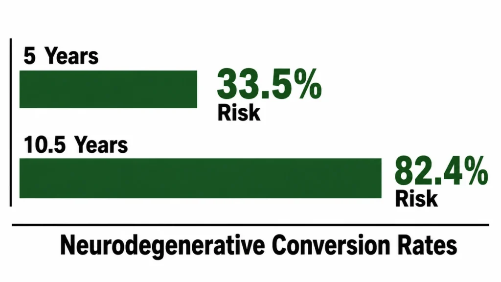
In the largest multicentre study of this kind, drawing on 1,280 polysomnography-confirmed cases across 24 international sleep centers, the average annual conversion rate was 6.25 percent.⁹
Conversion rates [the percentage of people with a prodromal condition who go on to develop the full disease within a given period] of this magnitude have no equivalent in any other recognized early marker of neurodegeneration.
This is the real finding: the sleep stage that produces your dreams has become the most reliable window medicine has for seeing neurological disease arrive, years before any movement or memory symptom appears.
These are probabilistic figures, not predictions for any individual. A person with confirmed isolated REM sleep behavior disorder is not certain to develop neurodegeneration. The conversion research cannot yet tell one individual whether they belong to the minority who never convert.
What it does tell all of us: acting out dreams in sleep is not a harmless quirk. Treating it as one means letting the single best diagnostic window close without looking through it.
Twelve years.
The average lag between REM sleep behavior disorder onset and first motor symptoms in one long-term cohort was 12 years.⁸ That is 12 years of possible monitoring, early planning, and clinical awareness.
The signs that deserve a real conversation with your doctor
Most people with REM sleep behavior disorder are not aware they have it. The movements happen while they are asleep, and a bed partner is often the first to notice. Many never present to medical attention, and the average delay from symptom onset to diagnosis is 7 to 9 years.⁴,⁹
That gap matters because the symptoms are specific. They are not the same as common nighttime twitches or the brief startle that happens as you fall asleep. The signs that warrant a clinical conversation look like this:
Talk to your doctor or ask for a sleep specialist referral if you or someone who shares your sleep space notices you are regularly doing any of the following during sleep:
- Punching, kicking, or flailing with force
- Shouting, screaming, or speaking in full sentences
- Sitting up, grabbing objects, or jumping from bed
- Acting out a storyline that matches a dream you recall when woken
- Behaviors that cluster in the second half of the night

Symptoms more likely to occur in the second half of the night set REM sleep behavior disorder apart from non-REM parasomnias, which tend to cluster in the first few hours.⁵
Diagnosis may require a sleep study called a polysomnogram [an overnight recording of brain activity, eye movements, muscle tone, and breathing during sleep] to confirm loss of REM atonia.⁴ Your primary care provider can make the referral; you do not need to see a neurologist first.
Before pursuing any treatment your doctor recommends for sleep-related symptoms, tell them about every medication you currently take, including antidepressants. Some selective serotonin reuptake inhibitors are associated with drug-induced REM sleep behavior disorder, and switching or adjusting them may be the first step.⁵
One practical starting point: keep a two-week sleep log noting any episodes described by a bed partner, then bring it to your appointment. This is exactly what sleep specialists ask for, and arriving with that record shortens the path to a useful evaluation.
What you can do right now, before any diagnosis exists
You do not need a diagnosis to start paying attention to this.
A 2021 review of management approaches found that higher physical activity is associated with a reduced risk of probable REM sleep behavior disorder, and researchers currently recommend exercise for all patients with the condition while neuroprotective medical therapies are still in development.¹⁰
Aerobic exercise [physical activity that raises your heart rate and keeps it elevated for a sustained period] shows the strongest association with protection across the broader synucleinopathy literature, though no clinical trial has yet confirmed it reduces conversion risk specifically in people with isolated REM sleep behavior disorder.¹⁰
For people who already have a confirmed diagnosis, the American Academy of Sleep Medicine recommends making the sleep environment physically safer while treatment decisions are made. This means:
- Moving sharp furniture or padding its edges near the bed
- Placing a soft mat on the floor beside the bed
- Removing glass objects, lamps, or weapons from the nightstand
- Considering bed rails or a sleeping bag to limit movement range⁵

Medications including immediate-release melatonin and clonazepam are conditionally recommended by the AASM for isolated REM sleep behavior disorder, with the choice depending on individual health factors and risk of side effects.⁵ These decisions belong with a clinician, not a supplement shelf.
The American Academy of Sleep Medicine’s patient resource site, Sleep Education (sleepeducation.org/find-a-sleep-center), lists accredited sleep centers by location and is freely accessible without a membership or paywall.
What you do with this information today may matter in ways that will only be measurable a decade from now.
The Window That Most People Never Open
REM sleep is not just a dream factory. It is a measurable window into the brainstem, and the behavior that happens during it, or fails to happen, is one of medicine’s most powerful early signals of neurological disease in adults over 50. If you or your bed partner have noticed kicking, shouting, or physically acting out dreams during sleep, bring a written description of those episodes to your next doctor’s appointment and ask whether a sleep study is appropriate. Who pays attention to this will matter.
⚠️DISCLAIMER
This article is for informational purposes only and does not constitute medical advice, diagnosis, or treatment. The content addresses recognizing symptoms of REM sleep behavior disorder and is intended for general educational purposes only. Health conditions vary significantly between individuals, always consult a licensed physician or qualified healthcare provider before making any decisions about your health or medical care.
References
- Van Cauter E, Leproult R, Plat L. Age-related changes in slow wave sleep and REM sleep and relationship with growth hormone and cortisol levels in healthy men. JAMA. 2000. https://pubmed.ncbi.nlm.nih.gov/10938176/
- Scarpelli S, Bartolacci C, D’Atri A, Gorgoni M, De Gennaro L. The functional role of dreaming in emotional processes. Frontiers in Psychology. 2019. https://pmc.ncbi.nlm.nih.gov/articles/PMC6428732/
- Zhang J, Pena A, Delano N, Sattari N, Shuster AE, Baker FC, Simon K, Mednick SC. Evidence of an active role of dreaming in emotional memory processing shows that we dream to forget. Scientific Reports. 2024. https://pubmed.ncbi.nlm.nih.gov/38622204/
- Roguski A, Rayment D, Whone AL, Jones MW, Rolinski M. A neurologist’s guide to REM sleep behavior disorder. Frontiers in Neurology. 2020;11:610. https://www.frontiersin.org/journals/neurology/articles/10.3389/fneur.2020.00610/full
- Howell M, Avidan AY, Foldvary-Schaefer N, et al. Management of REM sleep behavior disorder: an American Academy of Sleep Medicine clinical practice guideline. Journal of Clinical Sleep Medicine. 2023 https://pmc.ncbi.nlm.nih.gov/articles/PMC10071384/
- Takahashi R, Yamakado H, Uemura N, Taguchi T, Ueda J. The gut–brain axis based on α-synuclein propagation—clinical, neuropathological, and experimental evidence. International Journal of Molecular Sciences. 2025 https://pmc.ncbi.nlm.nih.gov/articles/PMC12072079/
- Dautan D, Paslawski W, Montejo SG, et al. Gut-initiated alpha synuclein fibrils drive parkinsonism phenotypes: temporal mapping of REM sleep behavior disorder-like and other non-motor symptoms. Translational Neurodegeneration. 2026. https://pmc.ncbi.nlm.nih.gov/articles/PMC12973632/
- Galbiati A, Verga L, Giora E, Zucconi M, Ferini-Strambi L. The risk of neurodegeneration in REM sleep behavior disorder: A systematic review and meta-analysis of longitudinal studies. Sleep Medicine Reviews. 2019;43:37-46. https://pubmed.ncbi.nlm.nih.gov/30503716/
- Postuma RB, Iranzo A, Hu M, et al. Risk and predictors of dementia and parkinsonism in idiopathic REM sleep behaviour disorder: a multicentre study. Brain. 2019;142(3):744-759. https://academic.oup.com/brain/article/142/3/744/5353011
- Matar E, McCarter SJ, St Louis EK, Lewis SJG. Current concepts and controversies in the management of REM sleep behavior disorder. Current Sleep Medicine Reports. 2021;7(1):19-32. https://pmc.ncbi.nlm.nih.gov/articles/PMC8116413/