

You’ve been checking your blood pressure. You’ve been watching your diet. But there’s something you probably haven’t looked at once since your last doctor’s visit.
Your own skin.
Most adults over 60 have no idea that their earlobes, shins, and nails are quietly displaying early signs of cardiovascular stress.
These signs appear on the outside of your body, in places you can see right now, before any chest pain or breathlessness begins. That’s the window this article is about.
The signs described in this article are potential risk indicators, not diagnostic tools. Only a qualified healthcare provider can evaluate your heart health through proper examination and testing. If you notice any of these signs, consult your doctor before drawing any conclusions.
The Earlobe Crease That Cardiologists Check First: What It Means for Your Heart After 60
You’ve probably looked at your earlobes a thousand times without seeing anything worth noting.
But a diagonal fold running across the earlobe, at roughly a 45-degree angle from the ear canal toward the outer edge, is something cardiologists have studied for decades.
It’s called Frank’s sign.
This crease is independently associated with cardiovascular risk. That means it shows up in people with heart disease more often than chance alone would explain, even when other risk factors are accounted for.¹

In a study of 805 adults, people over 55 were nearly five times more likely to have this crease than younger adults. People with acute coronary syndrome, such as a heart attack, were over five times more likely to have it.¹
That is a real signal. It doesn’t mean your heart is failing. It means your body may be displaying a cardiovascular risk marker in a place you can check without any equipment.
The crease matters more when it’s deep, complete across the full earlobe, and present on both ears. A faint line on one side is a lower-concern finding. A full, bilateral crease is worth mentioning to your doctor at your next visit.
Here’s the honest caveat. A 2023 European Heart Journal analysis found that while Frank’s sign is independently linked to coronary artery blockages, its diagnostic accuracy is modest.² It signals risk. It doesn’t confirm disease. Only a cardiologist with imaging and lab work can do that.
How to look at your earlobe

- Stand in good light near a mirror or take a close-up selfie.
- Pull the earlobe gently forward.
- Look for a diagonal line running from the small raised bump at your ear canal (the tragus) toward the outer edge of the lobe.
A faint or partial crease: note it and mention it at your next checkup.
A deep, full-width crease on both sides: document it with a photo and bring it up with your doctor within the week.
Any crease by itself is a flag, not a diagnosis.
The earlobe is one location. There are five more.
Shins That Stay Discolored or Won’t Heal: A Circulation Signal Hiding in Plain Sight
You look down at your lower legs and see reddish-brown skin. Maybe it’s been that way for months. You’ve called it a bruise, dry skin, or just something that comes with age.
It may be stasis dermatitis.
Stasis dermatitis is a form of inflammatory skin disease that typically affects the lower legs of older adults and represents a visible sign of venous hypertension.³
Venous hypertension means blood pressure inside the leg veins is too high because those veins can’t push blood back to your heart efficiently. The pressure builds, and it leaks through.
When that happens, red blood cells seep out of tiny vessels into the tissue just beneath the skin. They break down there. The iron they contain gets deposited in the skin.
That’s what causes the rust-colored discoloration you can see. The skin isn’t just stained. It’s inflamed.

This matters beyond skin care. The same vascular failure that causes this shin discoloration is also linked to reduced cardiac output and heart failure.
The veins and the heart are part of the same system. When one is struggling, it often means the other is, too.
One analysis of more than 4,000 geriatric patients found stasis dermatitis in 6.2% of people aged 65 and older.⁴ That’s more common than most people realize.
If left untreated, the inflammation can progress to open sores that are very slow to close. The skin doesn’t heal well once the circulation problem beneath it goes unaddressed.
You may have already seen this on your shins and dismissed it. That’s exactly the problem.
What the Color and Shape of Your Nails Are Telling a Cardiologist: Two Signs to Know
Most people think of nails as cosmetic. Cardiologists think of them differently.
Two nail findings are directly linked to cardiovascular disease, and both are visible without any tools. The first looks like something you’d dismiss immediately. The second looks like something that couldn’t possibly be serious.
That’s the problem.
Splinter hemorrhages are thin, dark lines running vertically under the nail. They look exactly like a wood splinter caught beneath the surface. Most of the time, a single line on one nail after some minor bumping is from trauma to the nail. That’s common and harmless.
But when multiple nails show these lines and there’s been no injury, it’s a different situation. Splinter hemorrhages appear in 15 to 33 percent of patients with infective endocarditis.⁵
Infective endocarditis is a bacterial infection of the heart valves. Small infected fragments break away from the valve, travel through the bloodstream, and lodge in the tiny vessels beneath your nails.
The line you see is damage from a clot that came from your heart.
One nail after a bump is almost certainly nothing. Several nails with no explanation is a reason to call your doctor.
Nail clubbing is the second sign. The fingertips widen and the nails curve downward, making the finger look broader at the tip.
Nail clubbing is most often associated with diseases of the heart and lungs, including cardiovascular disease.⁶ The body begins making these structural changes when oxygen delivery has been chronically low for a long time.
It’s not painful. It doesn’t announce itself. It builds slowly. And it’s visible every time you look at your hands.

The contradiction is this: both signs look cosmetic. Neither one feels alarming. That’s exactly why they get ignored.
Why Swollen Ankles and Shin Pitting Are Not Just Aging: They May Be Cardiac Edema
Someone in your life has probably told you that swollen ankles are just part of getting older. Maybe a doctor even said it. Maybe you said it to yourself.
That belief has a cost.
Ankle edema is a formal minor criterion in the Framingham Heart Study diagnostic framework for congestive heart failure.⁷
These criteria come from one of the longest-running cardiovascular studies in history. Ankle swelling has been on that list since 1971.⁷
That’s not “just aging.” That’s a documented cardiovascular signal.
Here is how cardiac edema works. When the heart’s pumping weakens, fluid backs up. Gravity pulls it downward into the legs. It starts at the ankles and moves up.
In a study of 279 acute heart failure patients, lower-leg edema was limited to the ankle in 22% of cases, reached the lower leg in 40%, and extended to the upper leg in 11%.⁸ The ankle-first, upward pattern is the cardiac pattern.
Pitting edema is the specific finding to check. Press your thumb firmly into your shin or the top of your foot for five seconds, then release.
If a dent stays visible for several seconds after you remove your thumb, that’s pitting edema. The tissue is waterlogged.

Cardiac edema also follows a time-of-day pattern. It typically worsens by the end of the day because you’ve been upright and gravity has been working.
It improves overnight after you’ve been lying flat. Swelling that follows this pattern, worsens progressively over weeks, and occurs in both legs deserves a cardiovascular evaluation.
Swelling from heat or long sitting can look similar. The difference is duration, progression, and the pitting test.
If you press your shin and a dent stays, don’t wait for your next routine appointment.
The Nail Finding That May Predict a Valve Problem Before Any Other Symptom Appears
You’ve noticed dark lines under a few of your nails. You haven’t hurt your hands recently. You search online and find nothing alarming.
This section is about what that search may have missed.
When splinter hemorrhages appear under multiple nails with no injury to explain them, they are potentially visible evidence of something happening at a heart valve.
Bacteria colonize the valve surface. The infection builds up material on the valve. Pieces break off. Those pieces travel through the bloodstream and reach the tiny capillaries beneath your nails.
The line you see under your nail is debris from a valve. It arrived through your bloodstream.
A 2024 peer-reviewed review in Open Medicine concluded that skin and nail lesions can act as significant clues in early diagnosis, helping prevent organ damage and allowing timely treatment.⁹
The point is that the external sign can appear while the internal damage is still developing. Treating the damage early changes the outcome.

Valve infections do not always cause pain. They don’t always cause a detectable murmur at first. They cause fatigue, fever, and sometimes nothing noticeable at all. But they can cause splinter hemorrhages. And those show up on your nails.
The cliffhanger here is not dramatic. It’s quiet. The nail finding appears first. The cardiac emergency comes later. Most people never connect the two.
If you see multiple unexplained dark lines under more than one nail, call your doctor that day. Don’t photograph them and file the photo away. Make the call.
What to Do When You Find One of These Signs: A Clear Action Sequence
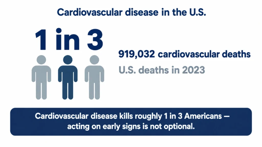
Heart disease remains the leading cause of death in the U.S. across men, women, and most racial and ethnic groups, with cardiovascular disease accounting for 919,032 deaths in 2023.¹⁰ That’s roughly one in every three deaths.
None of the signs in this article diagnose a heart condition. Only a doctor with tests can do that.
But the signs you’ve just read about are the kinds of things that appear before symptoms do. That’s the window.
When you find one of these signs, take three steps right away.
- Photograph it in good, natural light
- Write down when you first noticed it and whether it’s changed
- Note any other symptoms you’ve had: unusual fatigue, leg heaviness, breathlessness on stairs, or feeling your heart beating irregularly
Then contact your doctor. You don’t need to present a self-diagnosis. You need to say: “I noticed something I’d like you to look at in connection with my heart health.”
If you have more than one sign at once, don’t wait for a routine checkup.
Multiple findings together, an earlobe crease, shin discoloration, pitting ankles, or unexplained nail lines, raise the urgency. Request an expedited appointment and say why.
The danger is not the sign. The danger is seeing the sign and deciding it’s probably nothing.
Your body has been showing you information. Heart after 60 is a time when that information matters more, not less.
Acting on what you see is the difference between early intervention and a late one.
Check Your Body. Then Call Your Doctor.
Catching cardiovascular risk early is one of the most powerful things you can do for your heart after 60.
Schedule a cardiovascular screening and bring photos of any body changes to your appointment. Your body has already been trying to tell you something.
References
- Molina-Gallardo R, Aurelien-Cabezas NS, Tiburcio-Jimenez D, et al., Delgado-Enciso I. Traditional Cardiovascular Risk Factors Associated with Diagonal Earlobe Crease (Frank Sign) in Mexican Adults: Aging, Obesity, Arterial Hypertension, and Being Male Are the Most Important. International Journal of Hypertension. 2024; 2024:5598134. doi:10.1155/2024/5598134. https://www.ncbi.nlm.nih.gov/pmc/articles/PMC11213639/
- Gakovic B, Neskovic SA, Vranic I, Grujicic K, Ljubojevic A, Mijatovic S, Stankovic I. Diagonal earlobe crease (Frank’s sign) predicts obstructive coronary artery disease with modest accuracy. European Heart Journal. 2023; 44(Supplement 2):ehad655.1244. doi:10.1093/eurheartj/ehad655.1244. https://academic.oup.com/eurheartj/article/44/Supplement_2/ehad655.1244/7391308
- Silverberg J, Jackson JM, Kirsner RS, Adiri R, Friedman G, Gao XH, Billings SD, Kerkmann U. Narrative Review of the Pathogenesis of Stasis Dermatitis: An Inflammatory Skin Manifestation of Venous Hypertension. Dermatology and Therapy. 2023; 13:935-950. doi:10.1007/s13555-023-00908-0. https://link.springer.com/article/10.1007/s13555-023-00908-0
- Yalcin B, Tamer E, Toy GG, Oztas P, Hayran M, Alli N. The prevalence of skin diseases in the elderly: analysis of 4099 geriatric patients. International Journal of Dermatology. 2006; 45(6):672-6. (Cited through reference 3.)
- Manley A. Splinter haemorrhage. DermNet. 2019. https://dermnetnz.org/topics/splinter-haemorrhage
- Cleveland Clinic. Nail Clubbing. Cleveland Clinic Health Library. Last updated 23 November 2022. https://my.clevelandclinic.org/health/symptoms/24474-nail-clubbing
- Mahmood SS, Wang TJ. The epidemiology of congestive heart failure: the Framingham Heart Study perspective. Global Heart. 2013; 8(1):77-82. doi:10.1016/j.gheart.2012.12.006. https://pmc.ncbi.nlm.nih.gov/articles/PMC3756692
- Breidthardt T, Irfan A, Klima T, et al., Mueller C. Pathophysiology of lower extremity edema in acute heart failure revisited. American Journal of Medicine. 2012; 125(11):1124.e1-e8. doi:10.1016/j.amjmed.2011.12.015. https://pubmed.ncbi.nlm.nih.gov/22921885/
- Asif M, Yousuf MH, Farooqui US, Nashwan AJ, Ullah I. Cutaneous signs of selected cardiovascular disorders: A narrative review. Open Medicine. 2024; 19(1):20240897. doi:10.1515/med-2024-0897. https://www.ncbi.nlm.nih.gov/pmc/articles/PMC10921438/
- Centers for Disease Control and Prevention. Heart Disease Facts. Last updated 24 October 2024. https://www.cdc.gov/heart-disease/data-research/facts-stats/index.html